OSTEOSTRACI
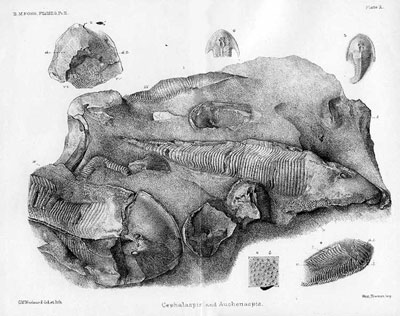
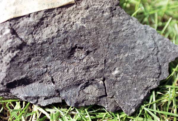
Catalogue of Fossil Fishes AS Woodward 1891 Cephalaspis murchisoni - Ledbury
The Osteostraci or Cephalaspids as they were once known are the most ‘famous’ of all fossil agnathans. The first forms are seen in the Wenlockian of Scotland (Ateleaspis) and the last (Escuminaspis) in the Frasian of Miguasha, Canada. They are thought to have lived in shallow marine/deltaic environments, but there is some suggestion that rivers may have played a part in their life-cycle. It is thought they were mostly suspension-feeders and may have been made extinct by the massive expansion of the antiarch placoderms which occupied the same niche in the mid-Devonian
They are identified by a large bony headshield containing the eyes, nasal
apparatus, pineal opening and the characteristic cephalic sensory fields at the
sides of the shield. Most Osteostraci have a horseshoe-shaped
headshield with sharp posterolateral cornual processes, to the inner margins of
which the pectoral fins are attached.
Osteostraci Classification
Osteostraci
Ateleaspis - Early Silurian (Wenlockian), Generalised form
Hirella
Hemicyclaspis
Cornuata
Cephalaspidida
Zenaspidida
Thyestiida
It has been suggested that most of the osteostracan diversication occurred before the Wenlockian because this period sees both Ateleaspis the most generalised primitive form and derived cornuates such as Tremataspis.
They are widely distributed across the Old Red Sandstone of Europe and North America. The UK has some spectacular sites.
Osteostraci in my collection (click on thumbnails to see larger images)
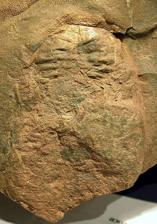
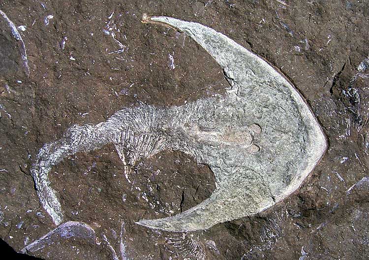
Ateleaspis tessellata traquair Silurian (Wenlockian) Lesmahagow/Hagshaw inlier, Scotland
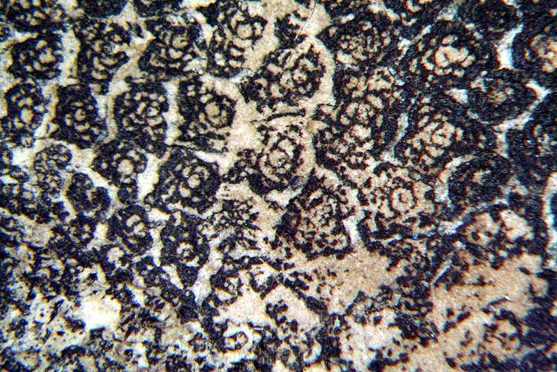
Ateleaspis is earliest vertebrate to have paired appendages. As with all Osteostraci, they also have a network of sensory fields made up of loose platelets of bone (not differentiated in ateleaspis) located along the mid-line and the lateral edges of the headshield. These sensory areas are connected to a complex of nerve channels and ducts which eventually connect with the labyrinth of the brain. It is thought that these area function as follows; when the animal moves up in the water the platelets are pushed down on the channels below them sending a signal to the portion of the brain associated with orientation and movement
In Ateleaspis, the sensory fields extend into the fins which are an extension of the headshield. Thus it has been suggested that the fins and fields developed together as means of stabilizing swimming along with the sense organs to provide feed-back control for it.


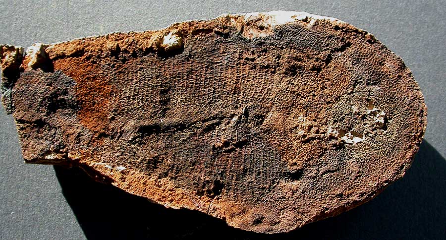
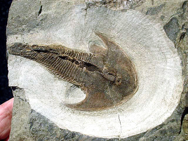
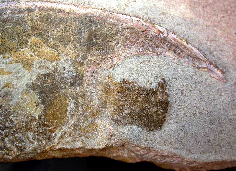
 An extraordinary complete dorsal fish with the head and body preserved in a
nodule and with the tail preserved in fish-bed. A true masterpiece of
fossil preparation.
An extraordinary complete dorsal fish with the head and body preserved in a
nodule and with the tail preserved in fish-bed. A true masterpiece of
fossil preparation.
![]()
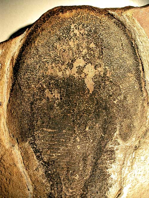
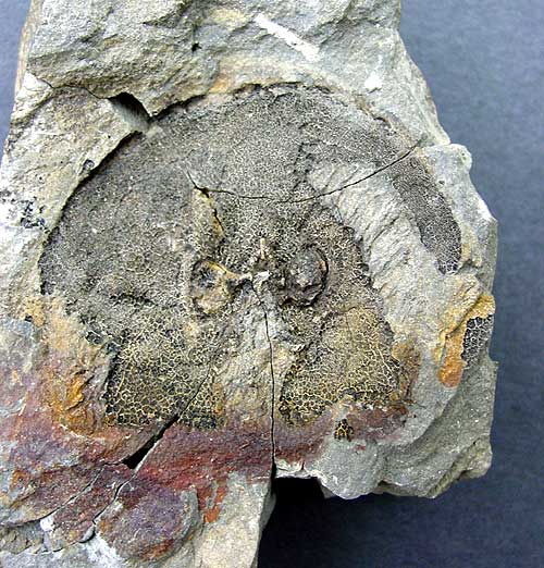
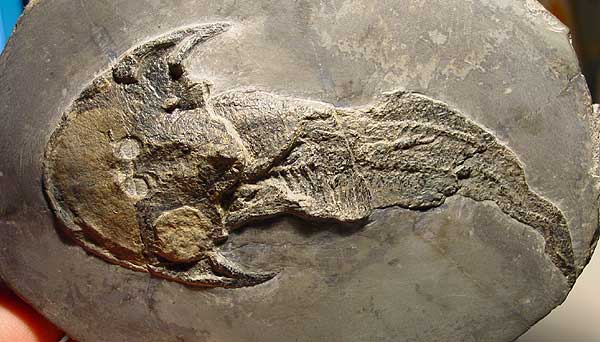
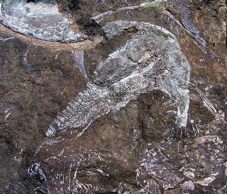
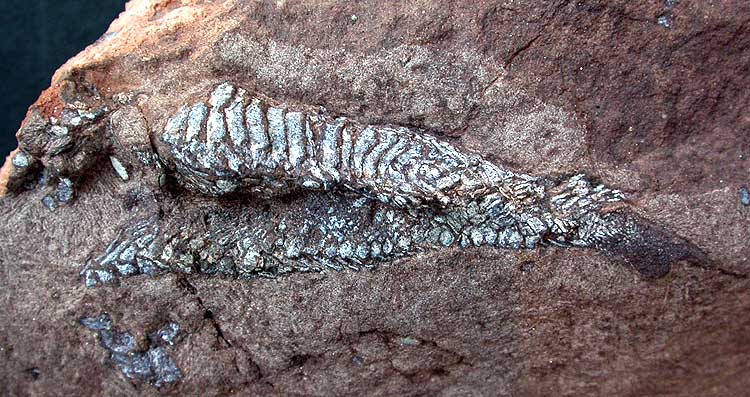
 An almost complete animal ventrally preserved in a nodule.
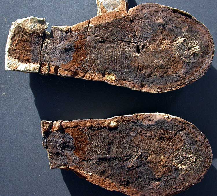
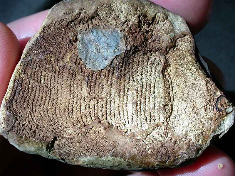
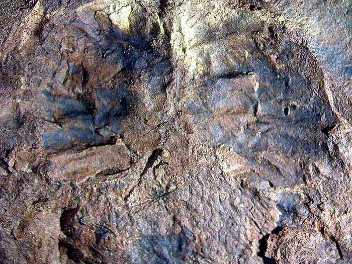
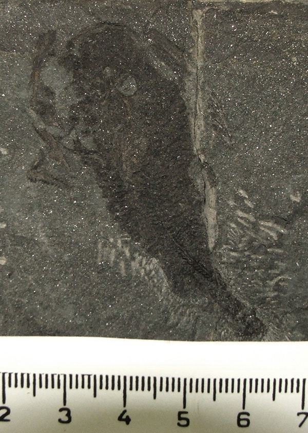
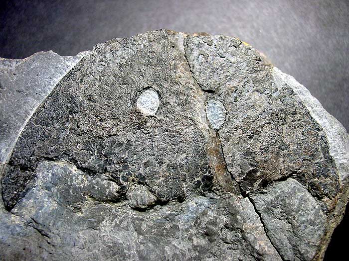
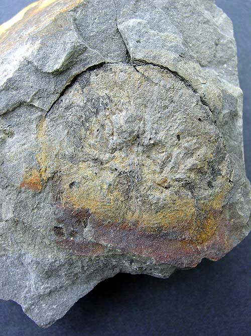 Pos/Neg of an Ateleaspis head preserved in fishbed

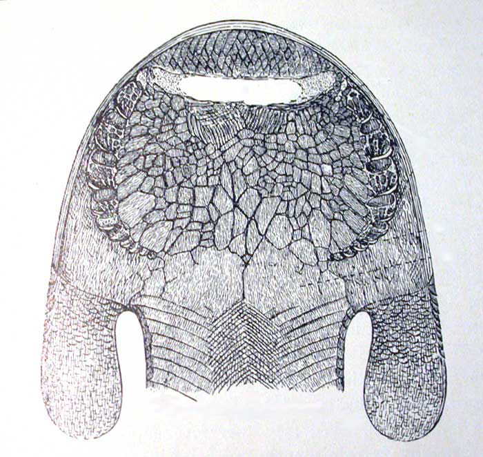
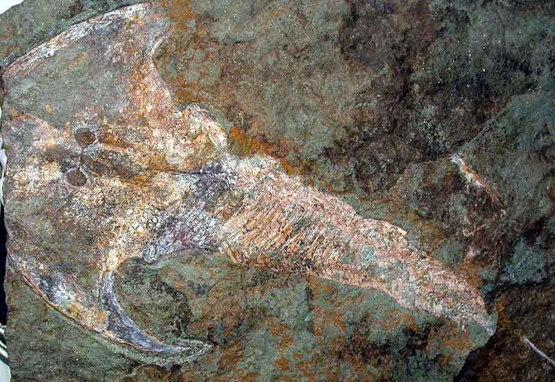
Hirella gracilis Upper Silurian Ringerike, Norway
Hirella is a basal osteostracan only marginally more complex than Ateleaspis. It has narrow paired fins and an oralobranchial chamber closed ventrally by large dermal platelets
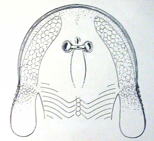 From Heintz 1939
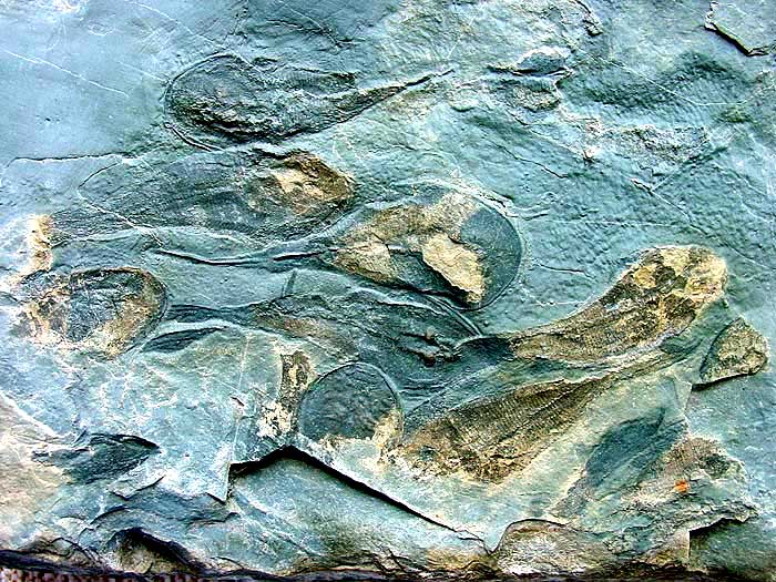
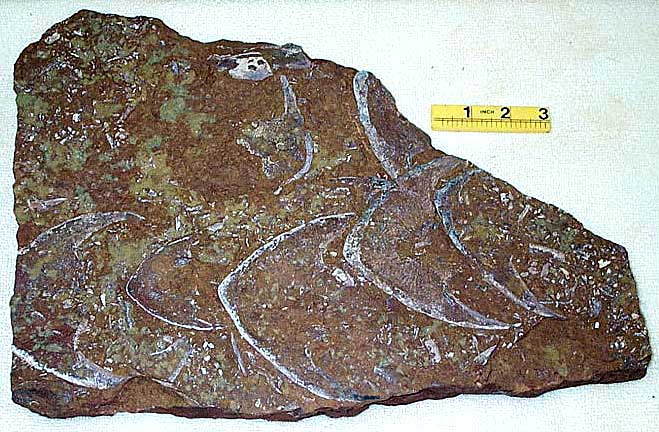
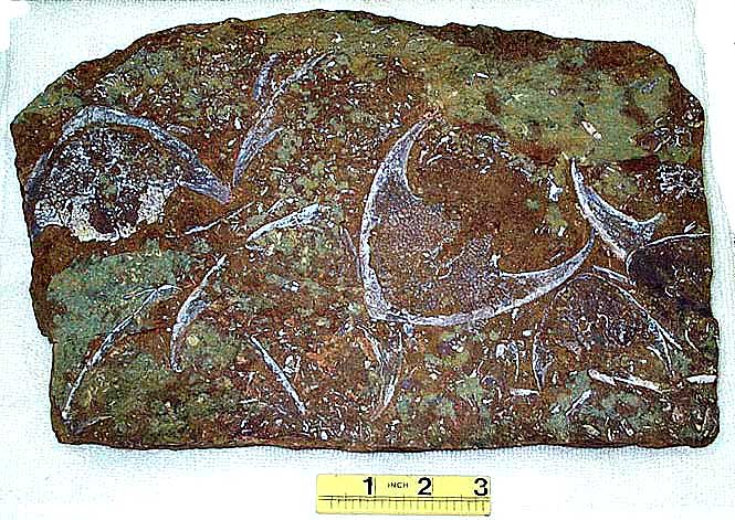
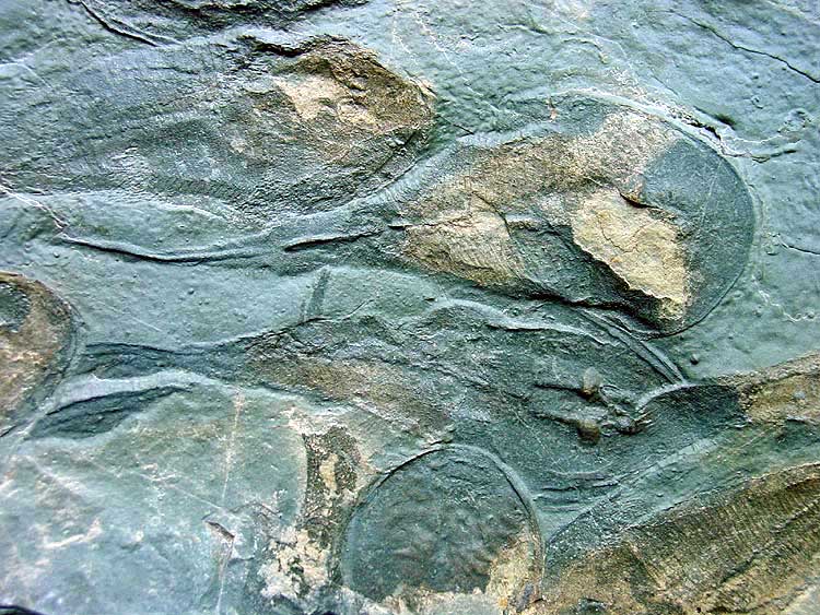 Mass-mortality plate

 Tail
Tail
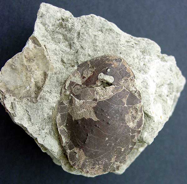
Zenaspi
dsThe Zenaspids are usually large osteostracans with tuberculated ornamentation and a large hypophyseal foramen. They are found in the Lower Devonian of Britain and Podolia They have a massive headshield with posteriorly enlarged sensory fields and thick, narrow cornual processes.
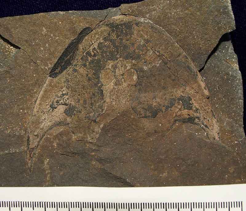
 Zenaspis pagei
Lower Devonian, Turin Hill, Scotland
Zenaspis pagei
Lower Devonian, Turin Hill, Scotland
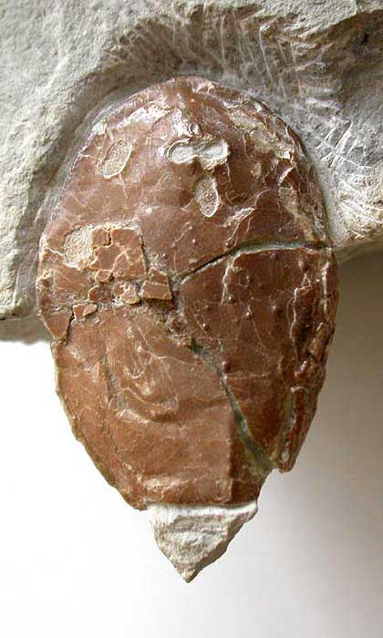
 Zenaspis powriei
Lower Devonian, Scotland
Zenaspis powriei
Lower Devonian, Scotland
Cephalaspis sp. x 2 Welsh borders (part of the Wonderblock...see also acanthodian)
 St Maughans Formation, LORS, Brecon, Wales
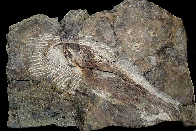
Yvonaspis campbelltonesis New Brunswick, Canada
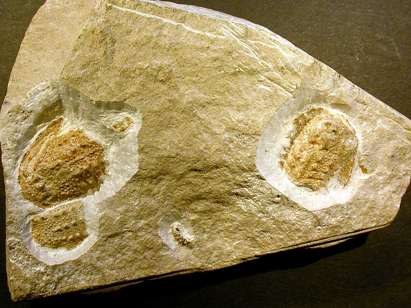
Osteostracan fauna from the Lower Devonian of Podolia, Ukraine
For the last 150 years, the Lower Devonian outcrops along the banks of the Dniester river in Podolia have been yielding wonderful fish remains. Despite the first descriptions of cephalaspids from the region being in 1874, little work has been done on the 19 species known, although recent papers by Janvier and Voichyshyn go a long way to addressing this lack of information.
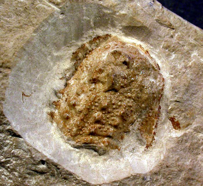
 Zychaspis siemiradzkii,
 Stensiopelta
pustulata
Stensiopelta
pustulata 
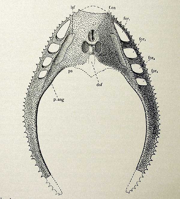
Z. siemiradzkii

![]()
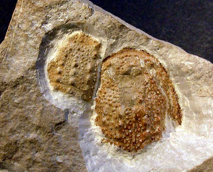
 The
ventral covering of the oralobranchial chamber, and one of the pectoral
The
ventral covering of the oralobranchial chamber, and one of the pectoral
fins
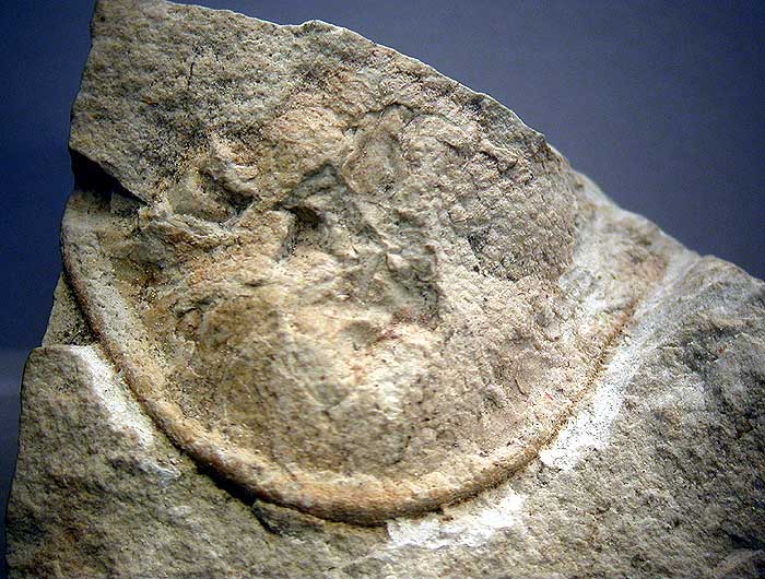
 horizontal ventral caudal lobe, a unique character of osteostracans that is
rarely observed
horizontal ventral caudal lobe, a unique character of osteostracans that is
rarely observed
 Z. siemiradzkii


Osteostracan fauna from the Silurian (Wenlock-Ludlow) Saaremaa Island, Estonia
The Osteostracans of Saarema island in Estonia, while being very early in the fossil record are a highly specialised group of small thyestiida. While most of this group show the classic cornuate morphology (Thyestes) others such as Witaaspis and Tremataspis are non-cornuate.
 Thyestes
 Witaaspis schrencki
![]() Tremataspis
mammillata
Tremataspis
mammillata
 Tremataspis schmidti
Osteostraci images from literature
 Poissons fossiles Agassiz Cephalaspis Lyelli
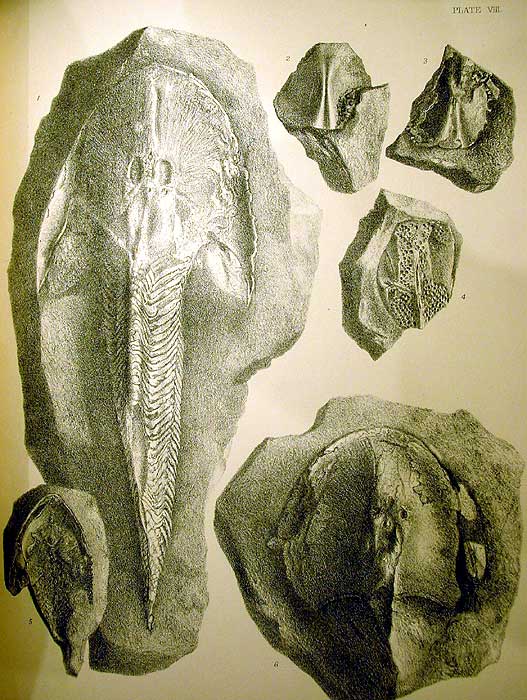
 Cephalaspids of Great Britain Stensio1932 Cephalaspis powriei Lankester
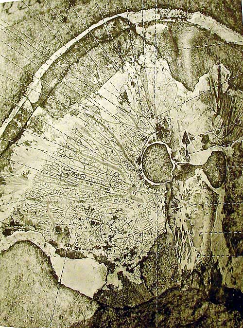 Stensio 1932 Cephalaspis whitei showing canals of sub-aponeurotic
vascular plexus
Stensio 1932 Cephalaspis whitei showing canals of sub-aponeurotic
vascular plexus
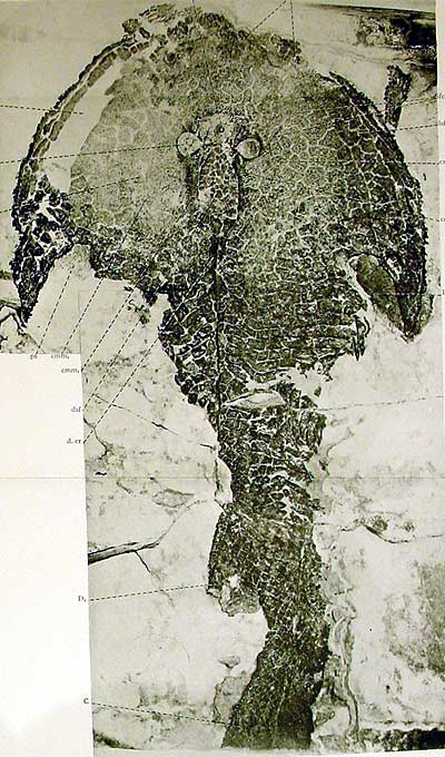
 Cephalaspids of Great Britain Stensio 1932 Cephalaspis pagei
 Cephalaspids of Great Britain Stensio 1932 Sclerodus pustuliferous agassiz
 Fishes of the Old Red Sandstone of Britain Powrie and Lankester 1868 (C.powrie
pagei asper)
Fishes of the Old Red Sandstone of Britain Powrie and Lankester 1868 (C.powrie
pagei asper)